15++ Pineal Gland Ct Brain
Pineal Gland Ct Brain. To get an accurate diagnosis, a piece of tumor tissue will be removed during surgery, if possible. About 1% of all brain tumors arise in the pineal gland.

Pineal gland tumor is a neoplasm that arises from the cells in pineal gland. Here are a number of highest rated pineal gland ct scan pictures upon internet. To get an accurate diagnosis, a piece of tumor tissue will be removed during surgery, if possible.
peinture rose poudre leroy merlin place des terreaux avant pate a eau persienne pvc blanc
Pineal Gland & Symbolism, page 1
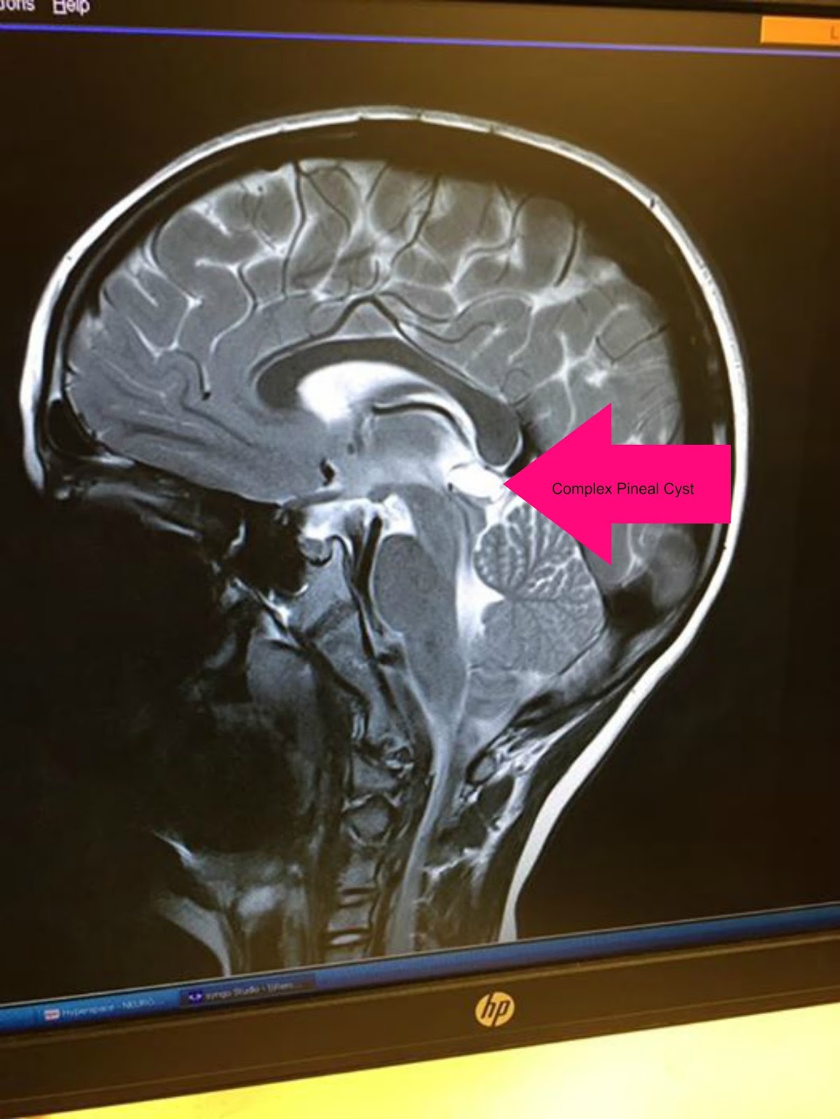
This is the hormone that regulates sleep. Sometimes an mri of the pineal cyst needs to be repeated with an intravenous contrast (dye) to rule out a pineal tumor. The base of the pineal stalk possesses a recess that is related with the third ventricle. The pineal gland is composed of pinealocytes and supporting cells that resemble the astrocytes present in the brain.

About 1% of all brain tumors arise in the pineal gland. A pineal tumor is a rare tumor of your pineal gland in your head. It functions in the secretion of melatonin. The pinealoma gland is a small gland found in the center of the brain. Ct of pineal region tumors the computed tomographic (ct) features of pineal region tumors.

It's surrounded by your brain. A pineal cyst seldom causes problems. In some cases, however, patients may complain of pain in head. Commonly calcified structures of the brain include the choroid plexus, pineal gland, basal ganglia and falx. It is attached by a stalk to the posterior wall of third ventricle.

The pinealoma gland is a small gland found in the center of the brain. This gland is a small structure deep within the brain. If a pineal cyst grows large, it may affect your vision. These tumors begin in the brain (in the pineal gland) but can spread to the spinal cord. Here are a number of highest rated pineal.

It's surrounded by your brain. Pineal region tumors are primary central nervous system (cns) tumors. Benign lesions tend to occur in adolescents and young adults whereas highly malignant lesions affect mostly children and adolescents. These tumors begin in the brain (in the pineal gland) but can spread to the spinal cord. Includes the ocular motor centre and pupillary control centre;

Includes the ocular motor centre and pupillary control centre; It functions in the secretion of melatonin. The pineal gland is composed of pinealocytes and supporting cells that resemble the astrocytes present in the brain. Usually, the cyst is not accompanied by any apparent clinical picture. At least 17 different types of tumors may occur in this region, and many are.

There are several structures in the brain which are considered normal if calcified. Pineal tumors are very rare tumors. The adrenergic nerves entering the pineal gland regulate its functions. The gland is a tiny gland in the middle of your head. If a pineal cyst grows large, it may affect your vision.

Pineal tumors are very rare tumors. The structures evaluated consisted of (a) the pineal gland, (b) the choroid plexus, (c) the habenula, (d) the basal ganglia, (e) the tentorium cerebelli, sagittal sinus and falx cerebri, (f) vessels and (g) lens and other structures which could be calcified. Above is the habenular commissure and below it is the posterior commissure. Includes.

At least 17 different types of tumors may occur in this region, and many are benign. Fluoride is one of the very few substances that can cross the 'blood brain barrier' destroying this important gland & keeping you from your full potential. A neuropathologist should then review the tumor tissue. The structures evaluated consisted of (a) the pineal gland, (b).

Here are a number of highest rated normal chest ct scan labeled pictures upon internet. If a pineal cyst grows large, it may affect your vision. These patients had a history of head trauma and their ct scan did not show any evidence of pathological findings. This is the largest reported series of histologically verified pineal region tumors studied with.