50++ Pineal Gland Calcification Ct Scan
Pineal Gland Calcification Ct Scan. There are several structures in the brain which are considered normal if calcified. Pineal calcification is a process whereby calcified concretions of hydroxyapatite develop;

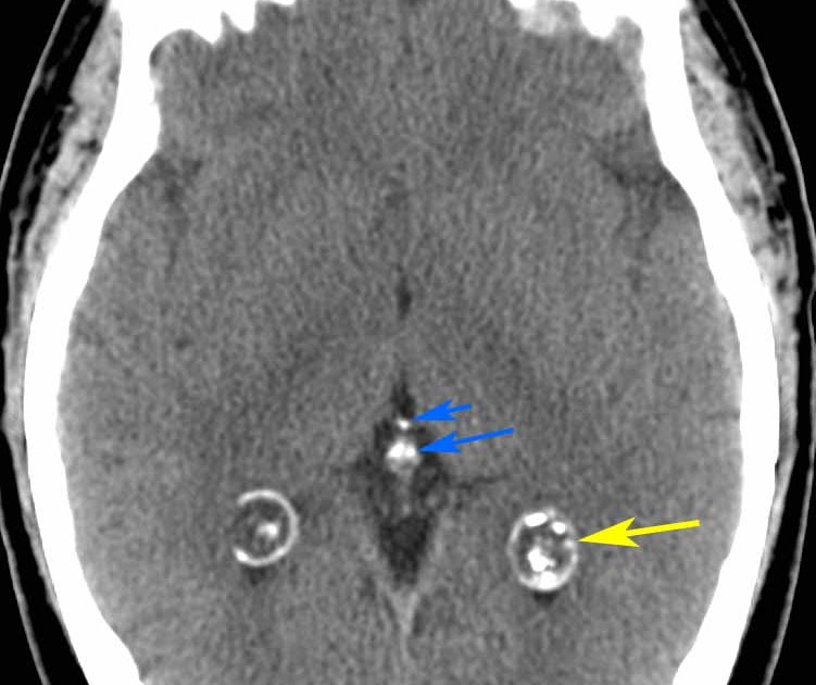
Commonly calcified structures of the brain include the choroid plexus, pineal gland, basal ganglia and falx. The mechanisms underlying the development of pineal calcification (pc) are elusive although there is experimental evidence that calcification may be a marker of the past secretory activity of the gland and/or of degeneration. Computed tomography studies concerning pineal calcification (pc) in schizophrenia have been conducted mainly by one author who correlated this calcification with several aspects of the illness.
papier peint motif rouge et gris pistolet a peinture bosch piscine pompe chaleur peinture pour escalier en bois brico depot
Pineal germinoma wikidoc
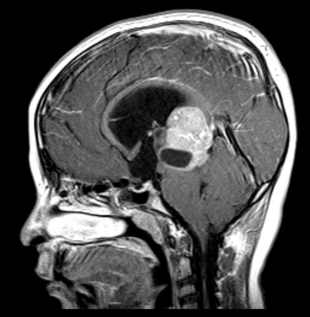
Pineal region tumors usually appear as a solid mass that brightens with contrast. There are several structures in the brain which are considered normal if calcified. Knowledge of these structures helps avoid confusion, especially when considering if. This calcification is observable by imaging techniques such as computed tomography (ct).

Germ cell tumors can be subdivided into two categories: The human pineal gland is a small neuroendocrine organ which produces melatonin.the main goal of this study was to provide a reference range for pineal volume in all age groups and to determine calcified and noncalcified tissue and their proportions, which may be a reflection of melatonin production in all age.

To investigate this relationship further, we studied in 47 ms patients (mean age: What do pineal region tumors look like on a ct scan or mri? Use of ct 'bone windows' is helpful in differentiating calcified structures from acute haemorrhage. Ad learn the shocking conditions that can be diagnosed with a ct scan right now. Calcification is thought to be.

We say you will this kind of pineal gland ct scan graphic could possibly be the most trending subject behind we ration it in google improvement or facebook. 13.6 ± 12.6 yrs) the association between fatigue and incidence of pineal calcification (pc) on ct scan, which is thought to reflect past secretory activity of the gland. Knowledge of these structures.

Ct scans were obtained before and af1er enhancement Pineal region tumors usually appear as a solid mass that brightens with contrast. On the basis of these findings the aim of the present study was to analyze size and incidence of pineal gland calcification by ct in schizophrenics and healthy controls, and to. Discover the common purposes, risks, and side effects.

There were 44 males and 16 females. Computed tomography (ct) scanning may be needed to evaluate a calcified pineal gland that is associated with a pineal germinoma or tumor calcification associated with other neoplasms in the pineal. Axial view of a head computed tomography (ct) scan of pineal gland calcification in the very center of the brain fluoride is the.

Pineal region tumors usually appear as a solid mass that brightens with contrast. On the basis of these findings the aim of the present study was to analyze size and incidence of pineal gland calcification by ct in schizophrenics and healthy controls, and to. Cerebral atrophy, which can be demonstrated on ct scan, is a common feature of ms resulting.