36+ Pineal Gland Calcification Ct
Pineal Gland Calcification Ct. Pineal calcification is a process whereby calcified concretions of hydroxyapatite develop; On the basis of these findings the aim of the present study was to analyze size and incidence of pineal gland calcification by ct in schizophrenics and healthy.

Pineal calcification is a process whereby calcified concretions of hydroxyapatite develop; The pineal gland has a predilection for calcification which is invariably histologically present in adults but rarely seen below the age of 10 years 6. In male patients, 70% of cranial ggct.
pistolet peinture haute pression airless peinture maison exterieur maroc peinture appui de fenetre blanc placard coulissant cuisine leroy merlin
Intracranial Calcification in Cone Beam CT & Medical CT
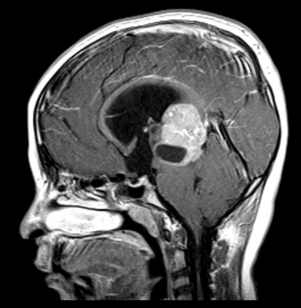
The mean age in the control and stroke groups were 58.18 and. Germ cell tumors can be subdivided into two categories: Compression of the quadrigeminal plate has caused obstructive hydrocephalus with corresponding ventricular dilation and transependymal csf flow. According to grech et.al, calcifications arising from the pineal gland itself often exhibit an exploded pattern whereas centrally located calcifications are produced by the pineal tumor (fig.11b).

The value of computed tomography (ct) for assessment of pineal calcifications was doi 10.1080/02841850600827585 # 2006 taylor & francis In male patients, 70% of cranial ggct. The incidence of normal pineal gland and choroids plexus calcification were higher in males than in females by 13.1% and 6.0% respectively. This calcification is observable by imaging techniques such as computed tomography (ct)..

To evaluate the prevalence of physiologic pineal calcification, estimate observer variability, and examine the association with choroid plexus calcifi… On the basis of these findings the aim of the present study was to analyze size and incidence of pineal gland calcification by ct in schizophrenics and healthy. An analytical cross sectional single center study was conducted. Compression of the quadrigeminal.

Pineal gland calcification was assessed by ct scan in 179 patients with ischemic stroke and 177 hospital controls. Calcification of the pineal gland. To evaluate the prevalence of physiologic pineal calcification, estimate observer variability, and examine the association with choroid plexus calcifi… Pineal gland calcification confirmed by ct scan is associated with ischemic stroke This calcification is observable by imaging.
 Source: neuroradiologyteachingfiles.com
Compression of the quadrigeminal plate has caused obstructive hydrocephalus with corresponding ventricular dilation and transependymal csf flow. These diameters were found to differ according to sex and age. Use of ct 'bone windows' is helpful in differentiating calcified structures from acute haemorrhage. To evaluate the prevalence of physiologic pineal calcification, estimate observer variability, and examine the association with choroid plexus.

Pineal gland calcification confirmed by ct scan is associated with ischemic stroke (a) axial ct shows a hyperdense tumor in the pineal region with central calcifications. This calcification is observable by imaging techniques such as computed tomography (ct). Calcified pineal displaced superiorly by inferiorly lo cated tumor. According to grech et.al, calcifications arising from the pineal gland itself often exhibit.

(a) axial ct shows a hyperdense tumor in the pineal region with central calcifications. The pineal gland has a predilection for calcification which is invariably histologically present in adults but rarely seen below the age of 10 years 6. The over all incidence of normal pineal gland calcification was 72.0 % and that of choroid plexus 43.3 %. Sometimes, the.

In male patients, 70% of cranial ggct. Compression of the quadrigeminal plate has caused obstructive hydrocephalus with corresponding ventricular dilation and transependymal csf flow. Choroid plexus calcification was found to have a prevalence of 53.6 %. Commonly calcified structures of the brain include the choroid plexus, pineal gland, basal ganglia and falx. On the basis of these findings the aim.

13.6 ± 12.6 yrs) the association between fatigue and incidence of pineal calcification (pc) on ct scan, which is thought to reflect past secretory activity of the gland. Knowledge of these structures helps avoid confusion, especially. Use of ct 'bone windows' is helpful in differentiating calcified structures from acute haemorrhage. Pineal gland calcification was assessed by ct scan in 179.

Knowledge of these structures helps avoid confusion, especially. An analytical cross sectional single center study was conducted. The value of computed tomography (ct) for assessment of pineal calcifications was doi 10.1080/02841850600827585 # 2006 taylor & francis Sometimes, the pineal gland develops calcium spots, also known as calcification. The mean age in the control and stroke groups were 58.18 and.